Fluorescence microscopeUsed to study the absorption, transport, distribution, and localization of chemicals within cells. There are two forms of fluorescence produced by the measured object: self-luminescence. The fluorescence emitted directly after ultraviolet irradiation is called secondaryFluorescence。 After fluorescent dye treatment and ultraviolet irradiation, it can be emittedFluorescence。 Some substances in cells, such as chlorophyll, produce self-luminescence when exposed to ultraviolet light. Although other substances themselves do notFluorescence, but they can be used by fluorescent dyes or...FluorescenceAntibody staining is released twiceFluorescence。
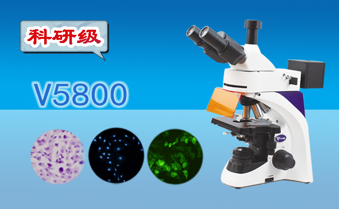
A fluorescence microscope selects a high-efficiency point light source and emits light of a certain wavelength (ultraviolet 365nm or violet-blue light 420nm) as excitation light. It excites the fluorescent substances in the specimen, emitting semi-transparent light of various colors, which is then observed through the magnification of objectives and eyepieces. In such a strong contrast environment, even faint translucent light is easy to recognize and has very high sensitivity. It is mainly used to study cell structure, function, and chemical composition.
Fluorescence microscopeThey can be divided into two types: scattering microscopy and descent microscopy. The former is primitive, the latter is advanced. The basic structures of the two types of fluorescence microscopes are similar, with the main differences as follows: scattered excited light passes through the specimen, generating light throughout the specimen, which then enters the objective lens. The greater the magnification, the weaker the light; Incident excitation light is projected onto the sample surface, generating light on the sample surface, which then enters the objective lens. The greater the magnification, the higher the light exposure, making it suitable for high-magnification observation.
Fluorescence microscopyThe main components of the mirror include mercury lamp sources, excitation filters, color separation mirrors (for detachment), suppression filters, and dark-field spotlights (scattering). Additionally, due to the severe heating of mercury lamps, most are equipped with heat-absorbing filters. Some fluorescence microscopes also have phase difference mirrors and annular appendix, allowing observation of phase differences. For fluorescence microscopes, choose inverted frames, inverted microscopes, etc.
In addition, all the aforementioned microscopes have microscope CCDs assembled into digital microscopes, which will be...MicroscopeThe physical images seen are converted into digital images and displayed on a computer. Therefore, we can study micro-domains through traditional binocular observation and reproduction on displays, thereby improving work efficiency.